Mummies and ancient DNA: Reading disease across Time
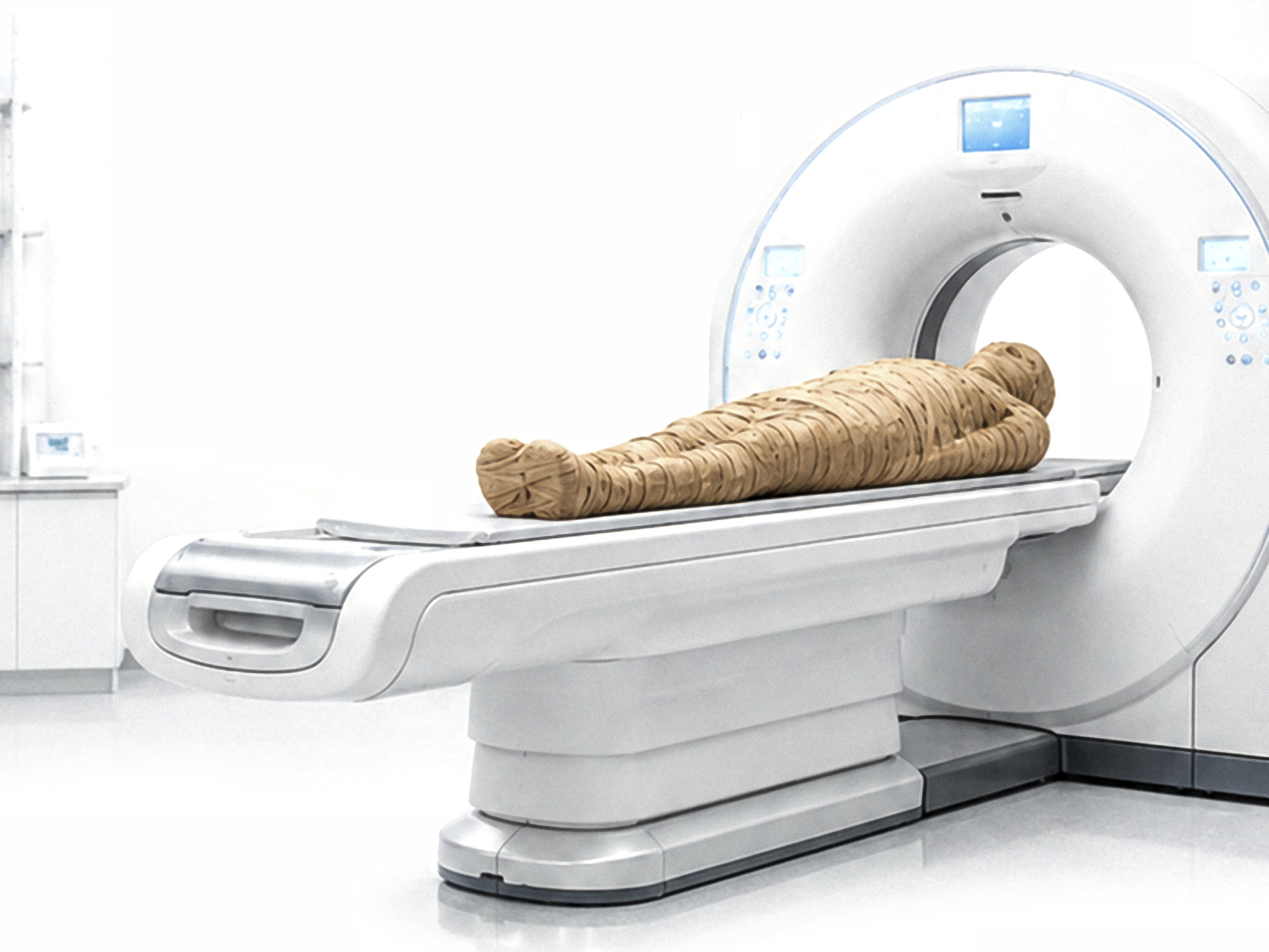
What can ancient human remains tell us about the evolution of health and disease? State-of-the-art imaging techniques and ancient pathogen DNA analysis can reveal hidden traces of disease, showing what biomedicine and archaeology can achieve together.
Mummies in the scanner: Insights into the past
Mummies are human bodies preserved for millennia. They are valuable for biomedical research but often fragile. State-of-the-art medical imaging allows us to look inside them without causing damage. X-rays, computed tomography and magnetic resonance imaging can reveal signs of disease, injury, ageing and mummification.
We show how researchers interpret these images, identify anatomical structures and signs of disease, and draw conclusions about life, disease, death and burial practices. By bringing together medicine, archaeology and anthropology, traces of the past become knowledge for the present.
Detecting ancient pathogen DNA
Ancient and historic human bodies may contain tiny fragments of pathogen DNA. These molecular traces can provide direct evidence of past infections and show how pathogens changed over time. For example, researchers can detect and analyse DNA from Mycobacterium leprae, the bacterium that causes leprosy. This helps us understand how infectious diseases affected earlier societies, how pathogens spread and evolved, and may offer insights relevant to present-day pandemics.
We explain why ancient DNA is fragile and easily contaminated, why samples must be studied in specialised clean-room laboratories, and how molecular data can answer archaeological, medical and evolutionary questions. Even tiny fragments can reveal new details about the history of infectious diseases and how humans and pathogens have interacted over time.